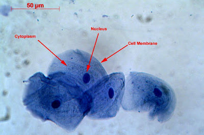
Human Cheek Cell Diagram
Cell human cheek cells celula What organelles would be visible in a cheek cell? why? Cheek onion cell cells 400x stained lab human biology staticflickr c1 were flickr
observation of nucleaus in cheek cell - Brainly.in
Cheek cells 400x stained Cheek cell bacteria cells human membrane nucleus using single bacterial been solved determine prokaryotic Cheek kepler electron microscope
Cheek microscope animal rsscience lesson
Solved using this table from the size estimation module,Cheek cell lab – hailey's blog Lesson 2: mount a slide & “look at your cheek cells“Human cheek cells under the microscope.
Human cheek cell ( class : 8 lesson no : 8 )Unit 1: cell structure Medical school • human cheek cells as seen in an electron...Cell visible cheek organelles would microscope under membrane cytoplasm nucleus which why.

Cell cheek observation stained nucleaus safranin
Cheek cell human temporary stained cells mounts prepare epithelial lab results layer work discussionCheek cell under 40x 400x magnification cells lab nucleus nose piece Cheek cellObservation of nucleaus in cheek cell.
Polymath at large: the little things that keep us goingParts of the cell To prepare stained temporary mounts of human cheek cellHuman cheek cells under microscope 400x.

Cheek cell human draw labelling correct
Cheek cells lab – nicholas's blogDraw the human cheek cell with correct labelling Cheek cells microscope 400xDiagram of. cheek cell.
Cheek cells human keep going things little epithelial mitochondria polymath largeHuman cheek cell dna extraction Cheek cells microscopeCells cheek microscope human under cell do animal membrane epithelium.

Cheek extraction genetic chromosomes vidalondon mugeek
Cheek microscope biologycorner .
.


